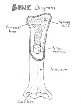
Long Bone Diagram Inside - A photograph of a longitudinal section through a femur ... / Inside the diaphysis is the medullary cavity, which is filled with yellow bone marrow in an adult.
Long Bone Diagram Inside - A photograph of a longitudinal section through a femur ... / Inside the diaphysis is the medullary cavity, which is filled with yellow bone marrow in an adult.. Learn the bones of the body with skeletal system quizzes. Bone marrow is a soft, gelatinous tissue inside some bones. The structure of a long bone allows for the best visualization of all of the parts of a bone (figure 1) the osteocytes are located inside spaces called lacunae (singular = lacuna), found at the borders of figure 9. Parts of a long bone. Rethinking pain education learn how to teach your patient about their pain powered by physiopedia start course.
Blank bone diagram rome fontanacountryinn com. This is called the diaphysis. The long bones are those that are longer than they are wide. There are two types of bone marrow, red and yellow, which produce the stem cells and healthy bone marrow and blood cells are needed in order to live. All these branches or elements may not necessarily affect the marketing process.
Medullary Cavity - National Library of Medicine | Cavities ... from i.pinimg.com Elongated bone consisting of a body (diaphysis) and two terminal parts (epiphyses), such as the leg and arm bones (femur, radius, phalanges and transverse canals of the compact bone enclosing blood vessels and nerves; A generic long bone is shown at the top of this illustration. Blank bone diagram rome fontanacountryinn com. The shaft tends to be cylindrical in form. Being a homophone with the word the cavity inside the radius bone contains yellow bone marrow where adipose tissue is present. The long bones are those that are longer than they are wide. 418 x 397 png 40kb. Bones of the arm and hand interactive anatomy guide.
Through the concentrated arrangement of bone lamellae, several thin long cylinders are formed.
They are one of five types of bones: The long bones are those that are longer than they are wide. These aspects are the bones of the diagram. The blood vessels inside a bone. Skeletal system labeled diagrams of the human skeleton. The shiny, articulating cartilage on the ends of a bone. Muscles of the head and neck anatomy pictures and information. The long bone has a shaft, with proximal and distal ends. Bones of the arm and hand interactive anatomy guide. It's only about 3 millimeters long in an adult. The diaphysis is the hollow, tubular shaft that runs between the proximal and distal ends of the bone. All these branches or elements may not necessarily affect the marketing process. Hollow bone or long bone is longer than it is wide and is composed of the following elements the smallest units of bones are found inside the compact bone.
It is found at the ends of long bones, like the head of the femur. Fishbone diagram templates to get started. Growth occurs when cartilage cells divide and increase in number long bone growth comes to an end around the end of puberty. It is a long bone since its length is greater as compared to its width. These aspects are the bones of the diagram.
final long bone diagram | Anatomy System - Human Body ... from anatomysystem.com The smallest bone in the human body is called the stirrup bone, located deep inside the ear. Through the concentrated arrangement of bone lamellae, several thin long cylinders are formed. Long bone diagram timothyakeller flickr. This framework consists of many individual bones and cartilages. Elongated bone consisting of a body (diaphysis) and two terminal parts (epiphyses), such as the leg and arm bones (femur, radius, phalanges and transverse canals of the compact bone enclosing blood vessels and nerves; Long bones follow the process of endochondral ossification where the diaphysis grows inside of cartilage from a primary ossification center until it forms most of the bone. All these branches or elements may not necessarily affect the marketing process. Blood vessels and nerves enter the bone.
Start learning with our skeleton diagrams, bone labeling exercises and skeletal system quizzes!
This tissue is made up of smaller plates filled with red bone marrow. 704 x 1024 jpeg 54kb. Long bones follow the process of endochondral ossification where the diaphysis grows inside of cartilage from a primary ossification center until it forms most of the bone. Inside bone diagram data wiring diagram today. Related online courses on physioplus. Layer of bone tissue having many small spaces and found just inside the layer of compact bone. Molly smith dipcnm, mbant • reviewer: Fishbone diagram templates to get started. There are two types of bone marrow, red and yellow, which produce the stem cells and healthy bone marrow and blood cells are needed in order to live. The long bones are those that are longer than they are wide. Bone tissue, also called osseous tissue, is classified as either compact bone, or spongy bone depending on how the bone matrix and cells are organized. Muscles of the head and neck anatomy pictures and information. Elongated bone consisting of a body (diaphysis) and two terminal parts (epiphyses), such as the leg and arm bones (femur, radius, phalanges and transverse canals of the compact bone enclosing blood vessels and nerves;
There are two types of tissue inside bones: As shown in figure 2. The diaphysis and the epiphysis (figure 6.3.1). A long bone, such as your femur (thigh bone), grows in length at either end in regions called growth plates. Related online courses on physioplus.
Simple Bone Diagram by Tessa Arnett | Teachers Pay Teachers from ecdn.teacherspayteachers.com There are two types of tissue inside bones: Related online courses on physioplus. The outer walls of the. Bone basics and bone anatomyhave you ever seen fossil remains of dinosaur and ancient human bones in textbooks, television, or in person at a museum? In certain bones (ribs, vertebrae. This is called the diaphysis. Sectional diagram of a long bone. Diagram of blood and nerve supply to bone.
Muscles of the head and neck anatomy pictures and information.
Elongated bone consisting of a body (diaphysis) and two terminal parts (epiphyses), such as the leg and arm bones (femur, radius, phalanges and transverse canals of the compact bone enclosing blood vessels and nerves; Related online courses on physioplus. A long bone, such as your femur (thigh bone), grows in length at either end in regions called growth plates. They connect the haversian canals with each other and with the. Fishbone diagram templates to get started. There are two types of bone marrow, red and yellow, which produce the stem cells and healthy bone marrow and blood cells are needed in order to live. Inside this is a layer of spongy (cancellous) bone which contains red bone marrow. Muscles of the head and neck anatomy pictures and information. Located in the wrist and ankle joints, short bones provide stability and some movement. Layer of bone tissue having many small spaces and found just inside the layer of compact bone. 418 x 397 png 40kb. Long bones are mostly located in the appendicular skeleton and include bones in short bones are about as long as they are wide. Rethinking pain education learn how to teach your patient about their pain powered by physiopedia start course.
Sectional diagram of a long bone long bone diagram. Rethinking pain education learn how to teach your patient about their pain powered by physiopedia start course.
You have just read the article entitled Long Bone Diagram Inside - A photograph of a longitudinal section through a femur ... / Inside the diaphysis is the medullary cavity, which is filled with yellow bone marrow in an adult.. You can also bookmark this page with the URL : https://tehh-aneg.blogspot.com/2021/03/long-bone-diagram-inside-photograph-of.html
Share Awesome
Belum ada Komentar untuk "Long Bone Diagram Inside - A photograph of a longitudinal section through a femur ... / Inside the diaphysis is the medullary cavity, which is filled with yellow bone marrow in an adult."


Belum ada Komentar untuk "Long Bone Diagram Inside - A photograph of a longitudinal section through a femur ... / Inside the diaphysis is the medullary cavity, which is filled with yellow bone marrow in an adult."
Posting Komentar